Article Science

Mapping cellular dynamics with the lichen cell atlas

Lichens thrive in the most extreme environments on earth; growing inside solid rock, and even surviving the vacuum of space. What can we learn from this fascinating and ancient partnership between two organisms?

14 July 2025

Lichens, believed to be one of the oldest forms of life on earth, are a symbiotic system consisting of a fungal partner and an algal (or, in some cases, cyanobacterial) partner. 

This partnership is based on the exchange of food in return for shelter. The alga uses photosynthesis to make food for the fungus, in return for a safe home. 

There are more than 20,000 known species of lichen, yet surprisingly function and evolution are still poorly understood.

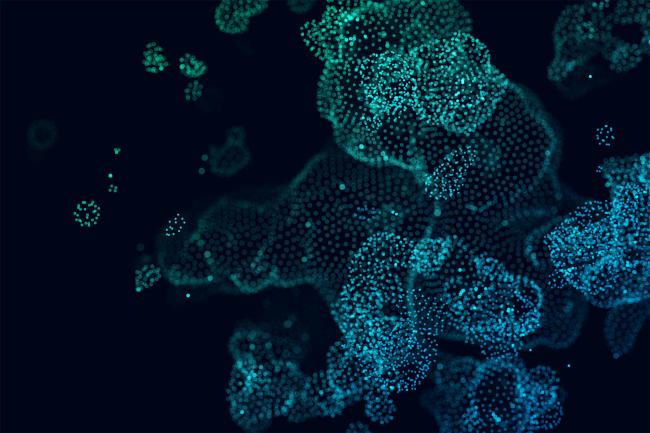
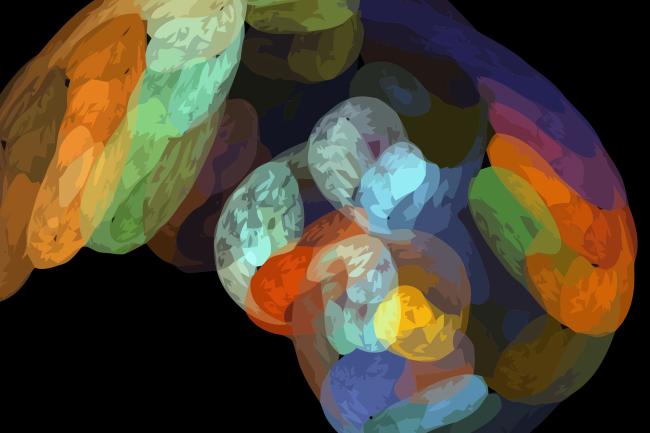
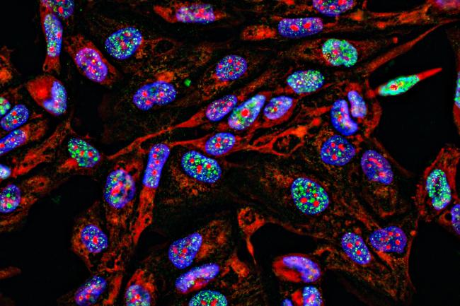
Dr Ellen Cameron, a postdoctoral researcher in the Papatheodorou group at the Earlham Institute, is developing a lichen cell atlas - a comprehensive and detailed map of all cell types in the organism. 

“We encounter lichens everywhere in our everyday lives,” she says. “They're globally distributed and ecologically significant, but often overlooked. Right now, I’m using single-cell transcriptomics to explore gene expression in lichens. 

“By looking for genes with specialised functions and different expression levels we can begin to disentangle how the fungi and algae interact and form these symbiotic associations. We can also begin to identify the diverse roles individual cells play within the lichen, which is very exciting. ”

Dr Cameron is working on common orange lichen, a common and widespread brightly coloured lichen growing in very diverse habitats.

Its fungal partner is Xanthoria parietina (the lichen is named after the fungal partner) and the algal partner is a unicellular green algae, a species of Trebouxia.

By looking for genes with specialised functions and different expression levels we can begin to disentangle how the fungi and algae interact and form these symbiotic associations.

Ellen Cameron presents her work on the Lichen Cell Atlas at a recent scientific seminar at EI

Dr Ellen Cameron presents her work on the Lichen Cell Atlas at this year's Science Symposium

Metagenomics to transcriptomics

There is relatively little transcriptomic work on lichens, and what does exist is typically bulk RNA sequencing. 

“Bulk sequencing is basically like a smoothie of all the gene expression data. One of the fun and challenging parts of my work is figuring out what to look for - our goal is to see whether the signals picked up in those bulk experiments are also visible at the single-cell level," Dr Cameron explains.

Dr Cameron’s background is in performing metagenomic assembly - the computational process of reconstructing complete or near-complete genomes from DNA sequences - to generate a reference genome.

“Having a reference genome is essential for mapping the single-cell transcriptomic data and identifying which genes are being expressed.

"When it comes to our single cell data, it is much more complex as it contains multiple species which we have a very limited understanding about. Working with non-model organisms in complex datasets like this doesn’t fit neatly into existing pipelines yet. So we’ve had to find the best way to analyze these datasets," she adds.

Working with non-model organisms in complex datasets like this doesn’t fit neatly into existing pipelines yet. So we’ve had to find the best way to analyse these datasets

Microscopic image of microalgae cells

Unicellular green algae, the algal partner.

Identifying interactions

Dr Cameron’s results distinguish between the transcriptomes of the fungal and algal partners - a really exciting development which demonstrates how single-cell transcriptomics can be applied in complex biological systems.

Looking to the future, her plan is to extend her work on the common orange lichen into multiple species.

“I’d love to expand this into a broader study involving multiple lichen species, to explore their evolution. Spatial transcriptomics would also allow us to confirm where in the lichen these different cell types are found.”

Her work generating the data was supported by EMBL-EBI and the Wellcome Sanger Institute. She is currently collaborating with Nick Talbot’s group at the Sainsbury Laboratory, generating single-cell data for their independent symbiont cultures to further understand the symbiosis in Xanthoria parietina

At the Earlham Institute, this project is part of the Institute’s strategic programme Cellular Genomics, which sheds light on how individual cells respond to their environment and contribute to the formation and maintenance of complex symbiotic relationships.

Keep up to date with our latest science

Image
Profile of Amy Lyall
Article author

Amy Lyall

Scientific Communications and Outreach Officer